- 翰林提供学术活动、国际课程、科研项目一站式留学背景提升服务!
- 400 888 0080
OCR A Level Biology:复习笔记2.2.11 Protein: Structure
Levels of Protein Structure
- There are four levels of structure in proteins, three are related to a single polypeptide chain and the fourth level relates to a protein that has two or more polypeptide chains
- Polypeptide or protein molecules can have anywhere from 3 amino acids (Glutathione) to more than 34,000 amino acids (Titan) bonded together in chains
Primary structure
- The sequence of amino acids bonded by covalent peptide bonds is the primary structure of a protein
- The DNA of a cell determines the primary structure of a protein by instructing the cell to add certain amino acids in specific quantities in a certain sequence. This affects the shape and therefore the function of the protein
- The primary structure is specific for each protein (one alteration in the sequence of amino acids can affect the function of the protein)
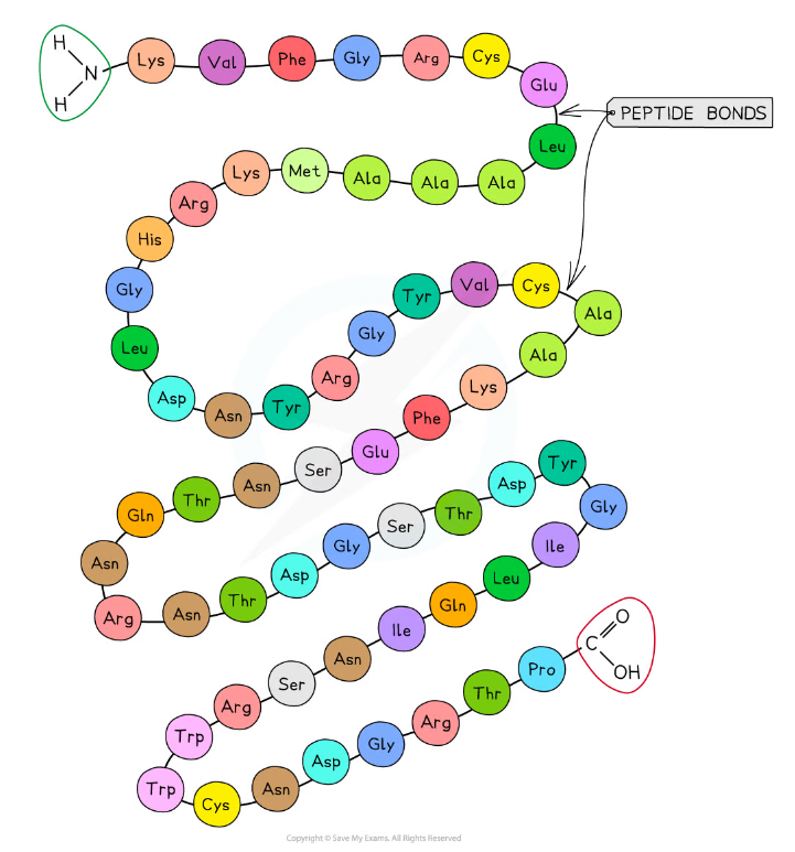
The primary structure of a protein. The three-letter abbreviations indicate the specific amino acid (there are 20 commonly found in cells of living organisms).
Secondary structure
- The secondary structure of a protein occurs when the weak negatively charged nitrogen and oxygen atoms interact with the weak positively charged hydrogen atoms to form hydrogen bonds
- There are two shapes that can form within proteins due to the hydrogen bonds:
- α-helix
- β-pleated sheet
- The α-helix shape occurs when the hydrogen bonds form between every fourth peptide bond (between the oxygen of the carboxyl group and the hydrogen of the amine group)
- The β-pleated sheet shape forms when the protein folds so that two parts of the polypeptide chain are parallel to each other enabling hydrogen bonds to form between parallel peptide bonds
- Most fibrous proteins have secondary structures (e.g. collagen and keratin)
- The secondary structure only relates to hydrogen bonds forming between the amino group and the carboxyl group (the ‘protein backbone’)
- The hydrogen bonds can be broken by high temperatures and pH changes
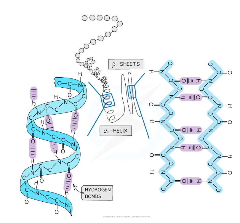
The secondary structure of a protein with the α-helix and β-pleated sheet shapes highlighted. The magnified regions illustrate how the hydrogen bonds form between the peptide bonds.
Tertiary structure
- Further conformational change of the secondary structure leads to additional bonds forming between the R groups (side chains)
- The additional bonds are:
- Hydrogen (these are between R groups)
- Disulphide (only occurs between cysteine amino acids)
- Ionic (occurs between charged R groups)
- Weak hydrophobic interactions (between non-polar R groups)
- This structure is common in globular proteins
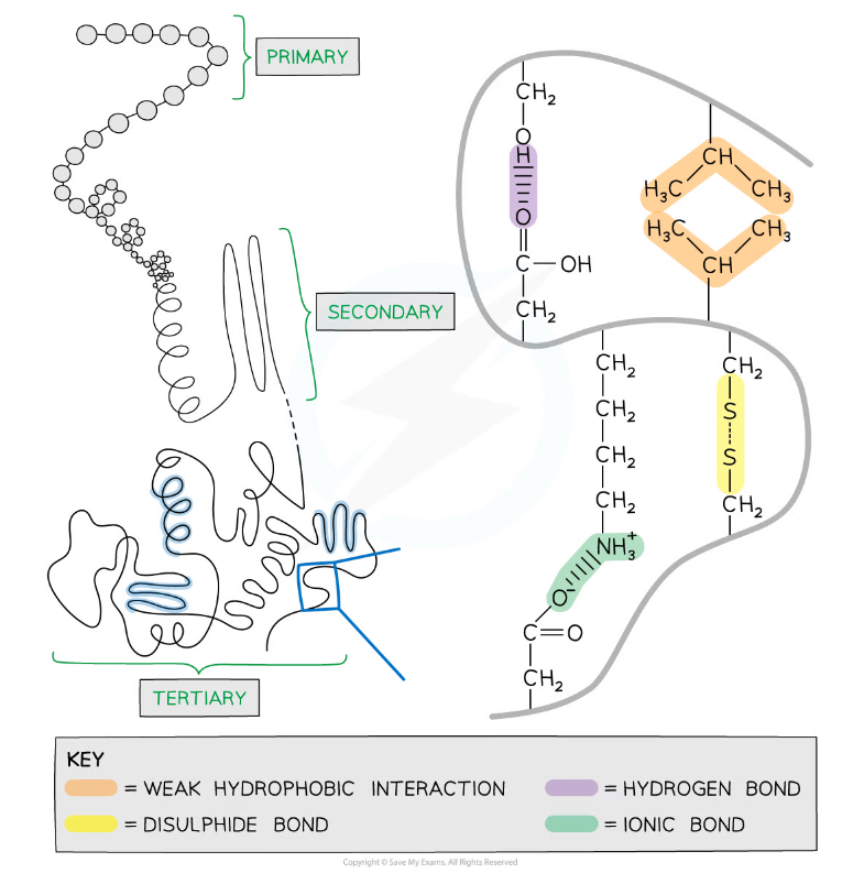
The tertiary structure of a protein with hydrogen bonds, ionic bonds, disulphide bonds and hydrophobic interactions formed between the R groups of the amino acids.
Disulphide
- Disulphide bonds are strong covalent bonds that form between two cysteine R groups (as this is the only amino acid with a sulphur atom)
- These bonds are the strongest within a protein but occur less frequently, and help stabilise the proteins
- These are also known as disulphide bridges
- Disulphide bonds can be broken by oxidation
- This type of bond is common in proteins secreted from cells eg. insulin
Ionic
- Ionic bonds form between positively charged (amine group -NH3+) and negatively charged (carboxylic acid -COO-) R groups
- Ionic bonds are stronger than hydrogen bonds but they are not common
- These bonds are broken by pH changes
Hydrogen
- Hydrogen bonds form between strongly polar R groups. These are the weakest bonds that form but the most common as they form between a wide variety of R groups
Hydrophobic interactions
- Hydrophobic interactions form between the non-polar (hydrophobic) R groups within the interior of proteins
Tertiary structure determines function
- A polypeptide chain will fold differently due to the interactions (and hence the bonds that form) between R groups
- Each of the twenty amino acids that make up proteins has a unique R group and therefore many different interactions can occur creating a vast range of protein configurations and therefore functions
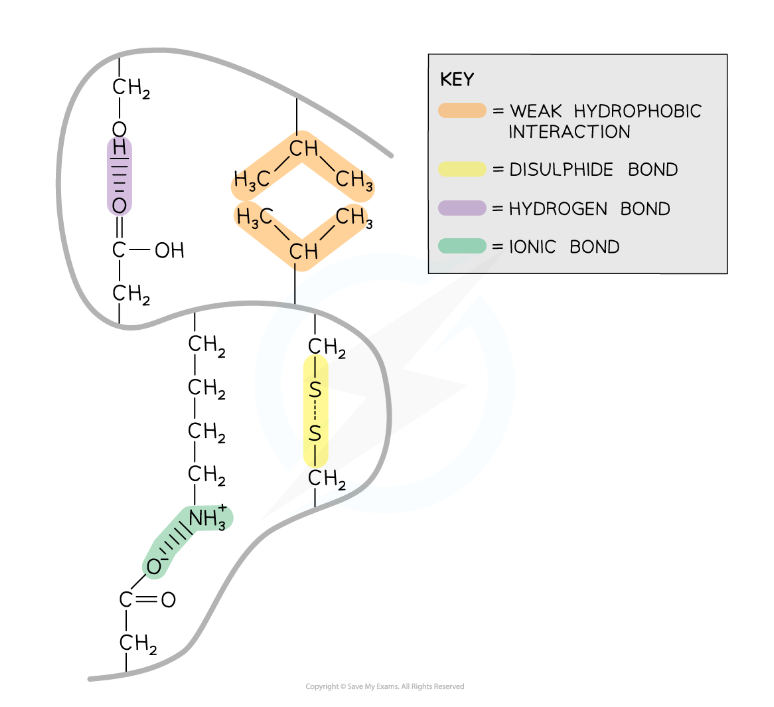
The interactions that occur between the R groups of amino acids determines the shape and function of a protein. These interactions are found within tertiary structures of proteins.
Quaternary structure
- Quarternary structure exists in proteins that have more than one polypeptide chain working together as a functional macromolecule, for example, haemoglobin
- Each polypeptide chain in the quaternary structure is referred to as a subunit of the protein
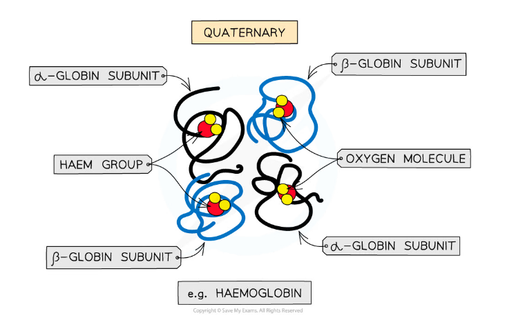
The quaternary structure of a protein. This is an example of haemoglobin which contains four subunits (polypeptide chains) working together to carry oxygen.
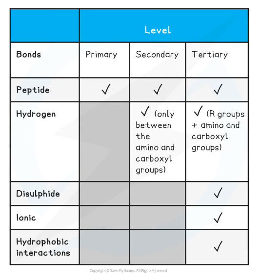
Summary of Bonds in Proteins Table
Exam Tip
Familiarise yourself with the difference between the four structural levels found in proteins, noting which bonds are found at which level. Remember that the hydrogen bonds in tertiary structures are between the R groups whereas in secondary structures the hydrogen bonds form between the amino and carboxyl groups.
转载自savemyexams

最新发布
© 2026. All Rights Reserved. 沪ICP备2023009024号-1


