- 翰林提供学术活动、国际课程、科研项目一站式留学背景提升服务!
- 400 888 0080
IB DP Biology: SL复习笔记6.2.4 The Blood System: Cardiac Cycle
Introduction to the Heart
Mammalian heart structure
- The heart is a hollow, muscular organ located in the chest cavity
- The heart is divided into four chambers
- The two top chambers are atria
- The bottom two chambers are ventricles
- The left and right sides of the heart are separated by a wall of muscular tissue called the septum.
- The septum is very important for ensuring blood doesn’t mix between the left and right sides of the heart
- Valves are important for keeping blood flowing forward in the right direction and stopping it flowing backwards. They are also important for maintaining the correct pressure in the chambers of the heart
- The atria and ventricles are separated by the atrioventricular valves
- The ventricles and the arteries that leave the heart are separated by semi-lunar valves
- The right ventricle and the pulmonary artery are separated by the pulmonary valve
- The left ventricle and aorta are separated by the aortic valve
- There are two blood vessels bringing blood to the heart; the vena cava and pulmonary vein
- There are two blood vessels taking blood away from the heart; the pulmonary artery and aorta
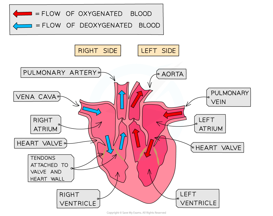
The human heart has four chambers and is separated into two halves by the septum
Sinoatrial Node
Contraction of the heart muscle
- The contraction of the heart is called systole, while the relaxation of the heart is called diastole
- Atrial systole is the period when the atria are contracting and ventricular systole is when the ventricles are contracting
- Atrial systole happens around 0.13 seconds after ventricular systole
- Atrial systole forces blood from the atria into the ventricles
- During ventricular systole, blood is forced from the ventricles into the pulmonary artery and aorta
- One systole and diastole makes a heartbeat and lasts around 0.8 seconds in humans.
- This is the cardiac cycle
Initiation of the heartbeat by the sinoatrial node
- The heart muscle is myogenic, meaning that the heart will beat without any external stimulus from other organs or the nervous system
- This intrinsic control causes the heart to beat at around 60 beats per minute
- The heart beat is initiated by a group of cells in the wall of the right atrium called the sinoatrial node (SAN)
- The cells of the sinoatrial node depolarise, reversing the charge across their membranes
- This triggers a wave of depolarisation that spreads across the rest of the heart
- The sinoatrial node is considered to be the pacemaker of the heart because it initiates the heart beat and so controls the speed at which the heart beats
- Note that artificial pacemakers are electronic devices implanted just underneath the skin. They can be used to replace or regulate the sinoatrial node if it becomes defective
Cardiac Cycle
Electrical control of the cardiac cycle
- The cardiac cycle is the series of events that take place in one heart beat, including atrial and ventricular systole
- The cardiac cycle is controlled by electrical signals that are initiated in the sinoatrial node
- Depolarisation of the cells in the sinoatrial node sends an electrical signal over the atria, causing them to contract in atrial systole
- The electrical signal then reaches a region of non-conducting tissue which prevents it from spreading straight to the ventricles; this causes the signal to pause for around 0.1 s
- This delay means that the atria can complete their contraction before the ventricles begin to contract
- The electrical signal is carried to the ventricles via the atrioventricular node (AVN)
- This is a region of conducting tissue between atria and ventricles
- The signal then travels to the base of the heart via conductive fibres in the septum known as the bundle of His
- The electrical signal is then carried through another set of conductive fibres called Purkyne fibres which spread around the sides of the ventricles, causing contraction of the ventricles from the apex, or base, of the heart upwards
- This is called ventricular systole
- Blood is forced out of the heart into the pulmonary artery and aorta
Electrical Control of the Cardiac Cycle Table
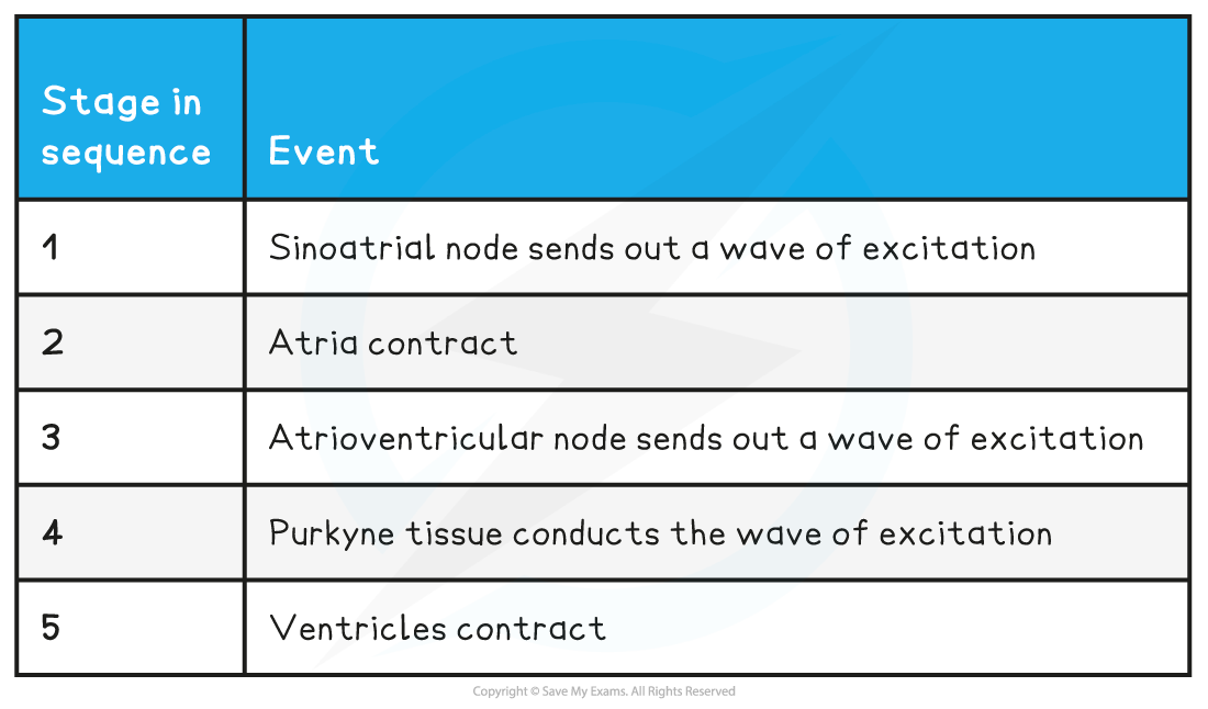
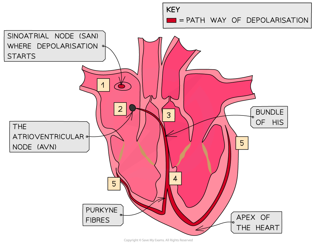
The wave of depolarisation spreads across the heart in a coordinated manner
Worked Example
Explain the roles of the sinoatrial node, the atrioventricular node and the conductive fibres in a heartbeat.
The cells of the sinoatrial node depolarise and send out an electrical signal which spreads across both atria, causing atrial systole. Non-conducting tissue between the atria and ventricles prevents depolarisation from spreading to the ventricles, ensuring that the atria finish contracting before the ventricles begin. The atrioventricular node then sends the electrical signal to the apex of the ventricles via conductive fibres in the septum known as the bundle of His. The electrical signal is then carried upwards around the walls of the ventricles by conductive tissues called Purkyne fibres. This means that during ventricular systole, the blood contracts from its base and blood is pushed upwards and outwards.
Cardiac Cycle Pressure Changes
- Contraction of the heart muscle causes an increase in pressure in the corresponding chamber if the heart, which then decreases again when the muscle relaxes
- Throughout the cardiac cycle, heart valves open and close as a result of pressure changes in different regions of the heart:
- Valves open when the pressure of blood behind them is greater than the pressure in front of them
- They close when the pressure of blood in front of them is greater than the pressure behind them
- Valves are an important mechanism to stop blood flowing backwards
- The pressure changes of the cardiac cycle can be represented in a graph
Pressure changes during diastole
- During diastole, the heart muscle is relaxing
- During this period, blood begins to flow into the atria from the veins, increasing the atrial pressure
- Relaxed muscle in the heart walls recoils, increasing the volume of the chambers of the heart:
- The atrioventricular valves open as the pressure in the atria is higher than the pressure in the ventricles
- The semilunar valves close as the pressure in the pulmonary artery and aorta is higher than the pressure in the ventricles
Pressure changes during systole
- During systole, the heart contracts and pushes blood out of the heart
- The contraction of the muscles in the wall of the heart reduces the volume of the heart chambers and increases the pressure within that chamber
- In atrial systole, the atria contract
- The atrioventricular valves are open as the pressure in the atria exceeds the pressure in the ventricles
- The semilunar valves are closed as the pressure in the ventricles is less than the pressure in the aorta and pulmonary artery
- In ventricular systole, the ventricles contract
-
- The atrioventricular valves are closed as the pressure in the ventricles exceeds the pressure in the atria
- The semilunar valves are open as the pressure in the ventricles exceeds the pressure in the aorta and pulmonary artery
-
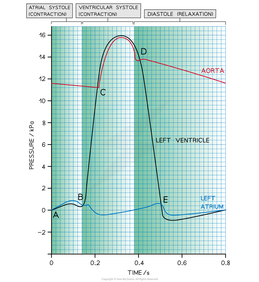
Graph showing the pressure changes within the aorta, left atrium and left ventricle during the cardiac cycle. The atrioventricular valves open at E and close at B, while the semilunar valves open at C and close at D.
Analysing the cardiac cycle
- The lines on the graph represent the pressure of the left atrium, aorta, and the left ventricle
- The points at which the lines cross each other are important because they indicate when valves open and close
Point A - the end of diastole
- The atrium has filled with blood during the preceding diastole
- Pressure is higher in the atrium than in the ventricle, so the AV valve is open
Between points A and B - atrial systole
- Left atrium contracts, causing an increase in atrial pressure and forcing blood into the left ventricle
- Ventricular pressure increases slightly as it fills with blood
- Pressure is higher in the atrium than in the ventricle, so the AV valve is open
Point B - beginning of ventricular systole
- Left ventricle contracts causing the ventricular pressure to increase
- Pressure in the left atrium drops as the muscle relaxes
- Pressure in the ventricle exceeds pressure in the atrium, so the AV valve shuts
Point C - ventricular systole
- The ventricle continues to contract
- Pressure in the left ventricle exceeds that in the aorta
- Aortic valve opens and blood is forced into the aorta
Point D - beginning of diastole
- Left ventricle has been emptied of blood
- Muscles in the walls of the left ventricle relax and pressure falls below that in the newly filled aorta
- Aortic valve closes
Between points D and E - early diastole
- The ventricle remains relaxed and ventricular pressure continues to decrease
- In the meantime, blood is flowing into the relaxed atrium from the pulmonary vein, causing an increase in pressure
Point E - diastole
- The relaxed left atrium fills with blood, causing the pressure in the atrium to exceed that in the newly emptied ventricle
- AV valve opens
After point E - late diastole
- There is a short period of time during which the left ventricle expands due to relaxing muscles
- This increases the internal volume of the left ventricle and decreases the ventricular pressure
- At the same time, blood is flowing slowly through the newly opened AV valve into the left ventricle, causing a brief decrease in pressure in the left atrium
- The pressure in both the atrium and ventricle then increases slowly as they continue to fill with blood
Exam Tip
When looking at the heart, remember the right side of the heart will appear on the page as being on the left. This is because the heart is labelled as if it were in your body and flipped around. Remember that the heart muscle is myogenic, which means that the heart will generate a heartbeat by itself and without any other stimulation. Instead, the electrical activity of the heart regulates the heart rate.The maximum pressure in the ventricles is substantially higher than in the atria. This is because there is much more muscle in the thick walls of the ventricles which can exert more force when they contract.
转载自savemyexams

早鸟钜惠!翰林2025暑期班课上线

最新发布
© 2025. All Rights Reserved. 沪ICP备2023009024号-1

